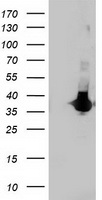
WB using E2F-1 pS364 ab shows detection of a ~47 kDa band corresponding to phosphorylated E2F-1 in induced cell lysates. Panel A shows reactivity using a control ab reactive to all forms of E2F (arrowheads). Panel B shows specific reactivity against phosphorylated E2F-1 (arrowheads) using anti-E2F-1 pS364. Lysates are as follows: CRE/E2F-1 are CRE cells derived from mouse NIH3T3 line transfected with human E2F-1, NIH-3T3 used as a negative control, and MDA-MB-231 cells are a human breast cancer line. Lysate was prepared from untreated cells or cells treated with 2 uM Doxorubicin used as DNA damaging agent. MDA-MB-231 cells were also treated with genistein, a mild DNA damaging agent. The figure shows the same membrane first probed with the anti-E2F-1 pS364 at 1:250, then stripped and re-probed with the pan E2F ab used as a positive control. The positive control ab shows an E2F-1 band in all human cell lines, but not mouse cells. Treatment with doxorubicin increases the expression of E2F-1 as shown in Panel A. After film development, images were overlapped to confirm that specific anti-E2F-1 pS364 staining shown treated human cells in Panel B specifically aligns with E2F-1 staining shown in Panel A.


