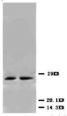
Figure 1. Immunoblot of Urm1 fusion protein: Anti-Urm1 antibody generated by immunization with recombinant yeast Urm1 was tested by immunoblot against yeast lysates expressing the Urm1-GFP fusion protein and other UBL fusion proteins. All UBLs possess limited homology to Ubiquitin and to each other, therefore it is important to know the degree of reactivity of each antibody against each UBL. Panel A shows total protein staining using ponceau. Panel B shows positions of free GFP or GFP containing recombinant proteins present in each lysate preparation after reaction with a 1:1,000 dilution of anti-GFP followed by reaction with a 1:15,000 dilution of HRP Donkey-a-Goat IgG. Panel C shows specific reaction with Urm1 using a 1:1,000 dilution of IgG fraction of Rabbit-anti-Urm1 (Yeast) followed by reaction with a 1:15,000 dilution of HRP Goat-a-Rabbit IgG. All primary antibodies were diluted in TTBS buffer supplemented with 5% non-fat milk and incubated with the membranes overnight at 4°C. Yeast lysate proteins were separated by SDS-PAGE using a 15% gel. This data indicates that anti-Urm1 is highly specific and does not cross react with other UBLs. Bands present in Panel C indicate that Urm1 and conjugated Urm1 is present in most yeast cell lysates albeit at significantly reduced levels relative to the Urm1-GFP transfected lysate. A chemiluminescence system was used for signal detection (Roche). Other detection systems will yield similar results. Data contributed by M. Malakhov, www.lifesensors.com, personal communication.


