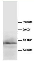
Figure. Immunoblot of SUMO-GFP fusion proteins cleaved by insect cell protein extracts. SUMO antibody is generated by immunization with recombinant Yeast SUMO, was tested by Immunoblot against several constructs of SUMO-GFP fusion proteins after cleavage by proteases in insect cell protein extracts. These constructs contained various linkers between the SUMO and GFP portion of the fusion proteins. Each sample was run twice. The left Lanes each contain 2ug E.coli expressed and purified SUMO-GFP fusion proteins after incubation with lysed cells (50ug total protein) for 1 h. The right Lanes contain the same fusion proteins incubated with the lysate in the presence of 2% SDS. After probing with anti-GFP antibodies the membranes were stripped of antibody using SDS-DTT solution for 30 mn at 60°C and were then re-probed using the SUMO antibody at a 1/1000 dilution incubated overnight at 4°C in 5% non-fat dry milk in TTBS. Detection occurred using a 1/2000 dilution of HRP-labeled Donkey anti-Rabbit IgG for 1 h at RT. A chemiluminescence system was used for signal detection (Roche). Other detection systems will yield similar results.


