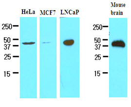
Cell lysates of HeLa, MCF7, LNCaP and mouse brain (30ug) were resolved by SDS-PAGE, transferred to NC membrane and probed with anti-human TREM2 (1:500). Proteins were visualized using a goat anti-mouse secondary antibody conjugated to HRP and an ECL detection system.

ICC/IF analysis of TREM2 in HepG2 cells line, stained with DAPI (Blue) for nucleus staining and monoclonal anti-human TREM2 antibody (1:100) with goat anti-mouse IgG-Alexa fluor 488 conjugate (Green).ICC/IF analysis of TREM2 in Raw264.7 cells line, stained with DAPI (Blue) for nucleus staining and monoclonal anti-human TREM2 antibody (1:100) with goat anti-mouse IgG-Alexa fluor 488 conjugate (Green).


