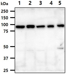
The cell and tissue lysates (40ug) were resolved by SDS-PAGE, transferred to PVDF membrane and probed with anti-human PPARGC1A antibody (1:1000). Proteins were visualized using a goat anti-mouse secondary antibody conjugated to HRP and an ECL detection system.Lane 1.: HeLa cell lysateLane 2.: MCF7 cell lysate Lane 3.: HepG2 cell lysate Lane 4.: 293T cell lysateLane 5.: Mouse brain tissue lysate

ICC/IF analysis of PPARGC1A in HeLa cells. The cell was stained with ATGA0466 (1:100). The secondary antibody (green) was used Alexa Fluor 488. DAPI was stained the cell nucleus (blue).


