Your Basket 0


Ordering Details

Cat Number:ATGA0240-50ul
Size:50uL
Concentration:1mg/mL
Price:£123.00
Quantity:
Shipping:£12.00

Document Links

MSDS


ACAT1 [AT15E5], Mouse Monoclonal Antibody

Partner: NKMax Bio (ATGen)

Applications:ELISA, WB, ICC/IF
Species Reactivity:Human
Isotype:Mouse IgG1 kappa
Immunogen:Recombinant human ACAT1 (34-427aa) purified from E. coli
Purification:By protein-A affinity chromatography
Conjugation:Unconjugated
Description:ACAT1 (acetyl-Coenzyme A acetyltransferase 1) is a 417 amino acid protein. ACAT1 is a mitochondrial enzyme involved in the formation and degradation of ketone bodies and is necessary for the proper metabolic processing of isoleucine. ACAT1 and ACAT2 catalyze the formation of acetoacetyl-CoA from two acetyl-CoA molecules. These enzymes are also capable of the reverse reaction. Defects in ACAT1 are a cause of 3-ketothiolase deficiency. 3-ketothiolase deficiency is an inborn error of isoleucine catabolism characterized by intermittent ketoacidotic attacks associated with unconsciousness. Some patients die during an attack or are mentally retarded.
Further Information:The antibody has been tested by ELISA, Western blot analysis to assure specificity and reactivity. Since application varies, however, each investigation should be titrated by the reagent to obtain optimal results. Recommended starting dilution is 1:1000.
Synonym:acetyl-CoA acetyltransferase 1, ACAT, MAT, T2, THIL
Ascension Number:P24752
NCBI EntrezGene:NP_000010
Shipping Conditions:Ship on cold packs
Storage:Can be stored at +2C to +8C for 1 week. For long term storage, aliquot and store at -20C to -80C. Avoid repeated freezing and thawing cycles.
Usage:Research use only


Insight Biotechnology Ltd reserves the right to change pricing without notice


The cell lysates (40ug) were resolved by SDS-PAGE, transferred to PVDF membrane and probed with anti-human ACAT1 antibody (1:1000). Proteins were visualized using a goat anti-mouse secondary antibody conjugated to HRP and an ECL detection system.The cell lysates (40ug) were resolved by SDS-PAGE, transferred to PVDF membrane and probed with anti-human ACAT1 antibody (1:1000). Proteins were visualized using a goat anti-mouse secondary antibody conjugated to HRP and an ECL detection system.Lane 1. : Hep3B cell lysateLane 2. : 293T cell lysateLane 3. : A431 cell lysate
The cell lysates (40ug) were resolved by SDS-PAGE, transferred to PVDF membrane and probed with anti-human ACAT1 antibody (1:1000). Proteins were visualized using a goat anti-mouse secondary antibody conjugated to HRP and an ECL detection system.The cell lysates (40ug) were resolved by SDS-PAGE, transferred to PVDF membrane and probed with anti-human ACAT1 antibody (1:1000). Proteins were visualized using a goat anti-mouse secondary antibody conjugated to HRP and an ECL detection system.Lane 1. : Hep3B cell lysateLane 2. : 293T cell lysateLane 3. : A431 cell lysate

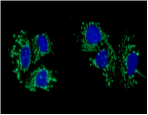
ICC/IF analysis of ACAT1 in Hep3B cells line, stained with DAPI (Blue) for nucleus staining and monoclonal anti-human   ACAT1 antibody (1:100) with goat anti-mouse IgG-Alexa fluor 488 conjugate (Green).

ICC/IF analysis of ACAT1 in Hep3B cells line, stained with DAPI (Blue) for nucleus staining and monoclonal anti-human ACAT1 antibody (1:100) with goat anti-mouse IgG-Alexa fluor 488 conjugate (Green).