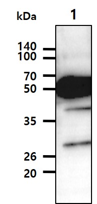
Cell lysates (35ug) were resolved by SDS-PAGE, transferred to PVDF membrane and probed with anti-human TUBB2b (1:500). Proteins were visualized using a goat anti-mouse secondary antibody conjugated to HRP and an ECL detection system.
Paraffin embedded sections of colorectal cancer tissue were incubated with anti-human TUBB2b antibody (1:50) for 2 hours at room temperature. Antigen retrieval was performed in 0.1M sodium citrate buffer and detected using Diaminobenzidine (DAB).

Immunofluorescence of human SW480 cells stained with monoclonal anti-human TUBB2b antibody (1:250) with Texas Red (Red). Arrow head: Mitotic cell, Arrow: MTOC (microtubule organizing center).


