Your Basket 0


Ordering Details

Cat Number:AP20619c
Size:400uL
Price:£304.00
Quantity:
Shipping:£12.00

Document Links

Data Sheet
MSDS


GLI2 [C-term] [RB49147], Rabbit Polyclonal Antibody

Partner: Abcepta, Inc.

Applications:IHC-P-Leica, IF, FC, WB
Species Reactivity:Human
Isotype:Rabbit Ig
Purification:Purified Rabbit Polyclonal Antibody
Description: Acts as a transcriptional activator. May play a role during embryogenesis. Binds to the DNA sequence 5'-GAACCACCCA-3' which is part of the TRE-2S regulatory element that augments the Tax-dependent enhancer of human T-cell leukemia virus type 1. Is involved in the smoothened (SHH) signaling pathway.
Further Information:This GLI2 antibody is generated from a rabbit immunized with a KLH conjugated synthetic peptide between 1287-1321 amino acids from the C-terminal region of human GLI2.
Synonym:Zinc finger protein GLI2, GLI family zinc finger protein 2 {ECO:0000312|HGNC:HGNC:4318}, Tax helper protein, GLI2 (<a href=http://www.genenames.org/cgi-bin/gene_symbol_report?hgnc_id=4318 target=_blank>HGNC:4318</a>)
Ascension Number:P10070
NCBI EntrezGene:2736
Shipping Conditions:Ship on cold packs
Storage:Maintain refrigerated at 2-8 C for up to 2 weeks. For long term storage store at -20 C in small aliquots to prevent freeze-thaw cycles.
Usage:Research use only


Insight Biotechnology Ltd reserves the right to change pricing without notice


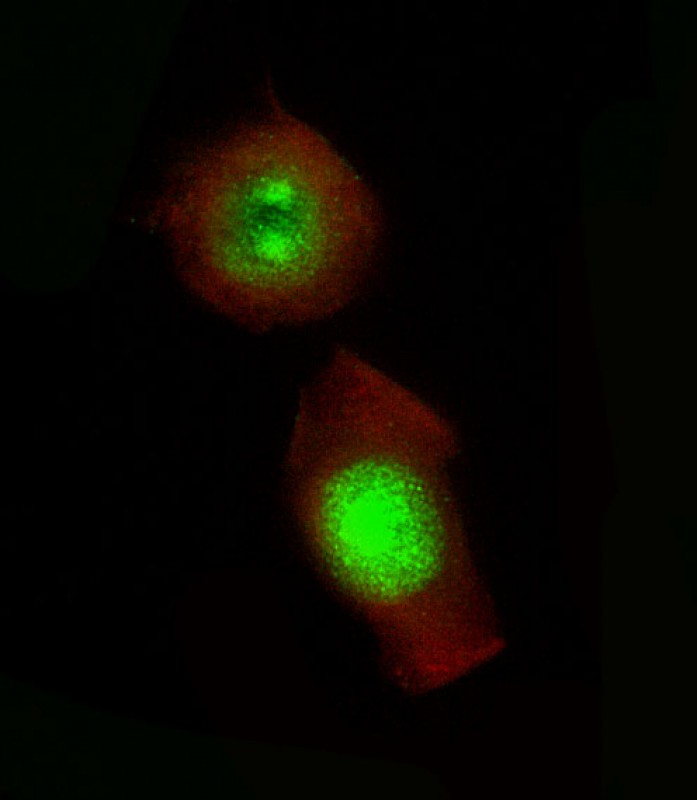
Immunofluorescent analysis of 4% paraformaldehyde-fixed,  0. 1% Triton X-100 permeabilized PC-3 cells labeling GLI2 with AP20619c at 1/25 dilution, followed by Dylight� 488-conjugated goat anti-Rabbit IgG secondary antibody at 1/200 dilution (green). Immunofluorescence image showing  Nucleus and Weak Cytoplasm staining on PC-3 cell line.  Cytoplasmic actin is detected with Dylight� 554 Phalloidin(red). The nuclear counter stain is DAPI (blue).

Immunofluorescent analysis of 4% paraformaldehyde-fixed, 0. 1% Triton X-100 permeabilized PC-3 cells labeling GLI2 with AP20619c at 1/25 dilution, followed by Dylight� 488-conjugated goat anti-Rabbit IgG secondary antibody at 1/200 dilution (green). Immunofluorescence image showing Nucleus and Weak Cytoplasm staining on PC-3 cell line. Cytoplasmic actin is detected with Dylight� 554 Phalloidin(red). The nuclear counter stain is DAPI (blue).