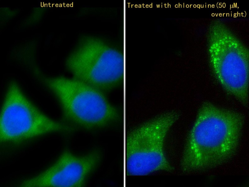
Immunofluorescent analysis of 4% paraformaldehyde-fixed, 0.1% Triton X-100 permeabilized Hela (human cervical epithelial adenocarcinoma cell line)(Hela-C:Serum-starve overnight;Hela-chloroquine?50 ?M, overnight; right) cells labeling GABARAP with AP1821a at 1/25 dilution, followed by Dylight� 488-conjugated goat anti-rabbit IgG (1583138) secondary antibody at 1/200 dilution (green). Immunofluorescence image showing cytoplasm and autophagic vacuoles staining on HeLa cell line. Cytoplasmic actin is detected with Dylight� 554 Phalloidin (PD18466410) at 1/100 dilution (red).The nuclear counter stain is DAPI (blue).


