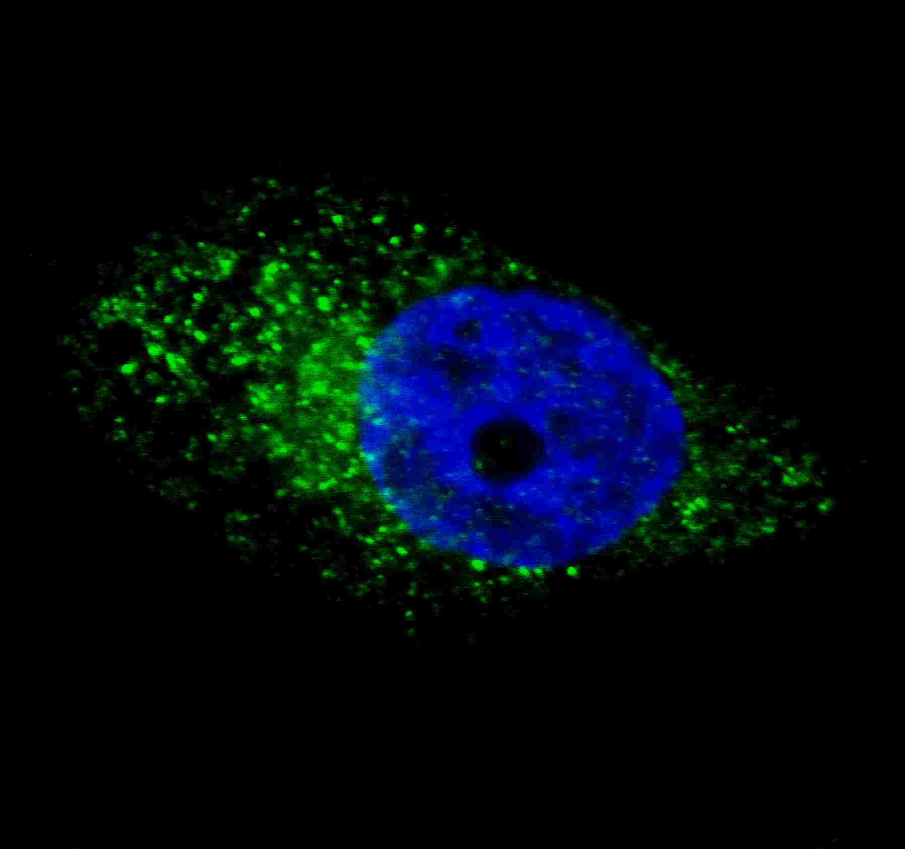
Fluorescent image of U251 cells stained with ATG16L antibody. U251 cells were treated with Chloroquine (50 ?M,16h), then fixed with 4% PFA (20 min), permeabilized with Triton X-100 (0.2%, 30 min). Cells were then incubated with AP1817d ATG16L primary antibody (1:100, 2 h at room temperature). For secondary antibody, Alexa Fluor� 488 conjugated donkey anti-rabbit antibody (green) was used (1:1000, 1h). Nuclei were counterstained with Hoechst 33342 (blue) (10 ?g/ml, 5 min). ATG16L immunoreactivity is localized to autophagic vacuoles in the cytoplasm of U251 cells, supported by Human Protein Atlas Data (http://www.proteinatlas.org/ENSG00000085978).


