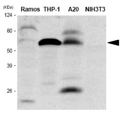
The extracts of Ramos, THP-1, A20 and NIH3T3 were resolved by SDS-PAGE, transferred to PVDF membrane and probed with anti-human IRF-5 antibody (1:1,000). Proteins were visualized using a goat anti-mouse secondary antibody conjugated to HRP and an ECL detection system.
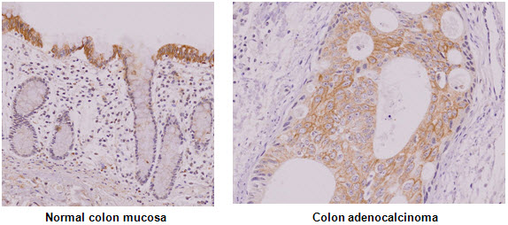
Paraffin embedded sections of normal colon mucosa and colon adenocalcinoma tissue were incubated with anti-human IRF-5 antibody (1:50) for 2 hours at room temperature. Antigen retrieval was performed in 0.1M sodium citrate buffer and detected using Diaminobenzidine (DAB).

ICC/IF analysis of IRF5 in THP-1 cells line, stained with DAPI (Blue) for nucleus staining and monoclonal anti-human THP-1 antibody (1:100) with goat anti-mouse IgG-Alexa fluor 488 conjugate (Green).ICC/IF analysis of IRF5 in Raw264.7 cells line, stained with DAPI (Blue) for nucleus staining and monoclonal anti-human IRF5 antibody (1:100) with goat anti-mouse IgG-Alexa fluor 488 conjugate (Green).


