Your Basket 0


Ordering Details

Cat Number:ABL1090-3
Size:200uL�5
Price:£759.00
Quantity:
Shipping:£12.00

Document Links

MSDS


Lamin B1 [15T1], Mouse Monoclonal Antibody

Partner: Abbkine Scientific

Applications:WB, IHC, IF, IP
Species Reactivity:Human, Mouse, Rat
Dilutions:Optimal working dilutions should be determined experimentally by the investigator. Suggested starting dilutions are as follows: WB (1:2000-1:5000), IHC-P (1:50-1:300), IF (1:200), IP (1:200).
Isotype:Mouse IgG
Immunogen:Recombinant Protein
Purification:The antibody was affinity-purified from mouse ascites by affinity-chromatography using specific immunogen
Description:Featured Anti-Lamin B1 Monoclonal Antibody (15T1), specially designed for your immunoassay as internal control.
Further Information:LMNB1 encodes one of the two B-type lamin proteins and is a component of the nuclear lamina. A duplication of LMNB1 is associated with autosomal dominant adult-onset leukodystrophy (ADLD). Alternative splicing results in multiple transcript variants.
NCBI EntrezGene:4001
Shipping Conditions:Ship on cold packs
Storage:Stable for one year at -20 C from date of shipment. For maximum recovery of product, centrifuge the original vial after thawing and prior to removing the cap. Aliquot to avoid repeated freezing and thawing.
Usage:Research use only


Insight Biotechnology Ltd reserves the right to change pricing without notice


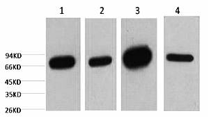
A01090-1.jpg&&Fig.1. Western blot analysis of 1) HepG2, 2) 293T, 3) mouse brain tissue, 4) rat brain tissue, diluted at 1:5000.|||A01090-2.jpg&&Fig.2 Immunofluorescence analysis of human lung cancer tissue. 1, Lamin B1 Monoclonal Antibody (15T1) (red) was diluted at 1:200 (4�C, overnight). 2, Cy3 Labeled secondary antibody was diluted at 1:300 (room temperature, 50min). 3, Picture B: DAPI (blue) 10min. Picture A: Target. Picture B: DAPI. Picture C: merge of A+B.|||A01090-3.jpg&&Fig.3. Immunohistochemical analysis of paraffin-embedded human uterus tissue. 1, Lamin B1 Monoclonal Antibody (15T1) was diluted at 1:200 (4�C, overnight). 2, Sodium citrate pH 6.0 was used for antibody retrieval (>98�C, 20min). 3, secondary antibody was diluted at 1:200 (room temperature, 30min). Negative control was used by secondary antibody only.

A01090-1.jpg&&Fig.1. Western blot analysis of 1) HepG2, 2) 293T, 3) mouse brain tissue, 4) rat brain tissue, diluted at 1:5000.|||A01090-2.jpg&&Fig.2 Immunofluorescence analysis of human lung cancer tissue. 1, Lamin B1 Monoclonal Antibody (15T1) (red) was diluted at 1:200 (4�C, overnight). 2, Cy3 Labeled secondary antibody was diluted at 1:300 (room temperature, 50min). 3, Picture B: DAPI (blue) 10min. Picture A: Target. Picture B: DAPI. Picture C: merge of A+B.|||A01090-3.jpg&&Fig.3. Immunohistochemical analysis of paraffin-embedded human uterus tissue. 1, Lamin B1 Monoclonal Antibody (15T1) was diluted at 1:200 (4�C, overnight). 2, Sodium citrate pH 6.0 was used for antibody retrieval (>98�C, 20min). 3, secondary antibody was diluted at 1:200 (room temperature, 30min). Negative control was used by secondary antibody only.