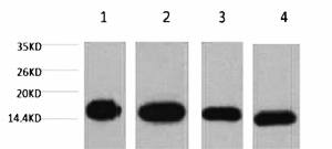
A01070-1.jpg&&Fig.1. Western blot analysis of 1) Hela, 2) Raw, 3) mouse brain tissue, 4) rat brain tissue, diluted at 1:5000.|||A01070-2.jpg&&Fig.2. Immunohistochemical analysis of paraffin-embedded human uterus tissue. 1, Histone H3 Monoclonal Antibody (2D10) was diluted at 1:200 (4�C, overnight). 2, Sodium citrate pH 6.0 was used for antibody retrieval (>98�C, 20min). 3, secondary antibody was diluted at 1:200 (room temperature, 30min). Negative control was used by secondary antibody only.|||A01070-3.jpg&&Fig.3. Immunohistochemical analysis of paraffin-embedded mouse testis tissue. 1, Histone H3 Monoclonal Antibody (2D10) was diluted at 1:200 (4�C, overnight). 2, Sodium citrate pH 6.0 was used for antibody retrieval (>98�C, 20min). 3, secondary antibody was diluted at 1:200 (room temperature, 30min). Negative control was used by secondary antibody only.|||A01070-4.jpg&&Fig.4. Immunohistochemical analysis of paraffin-embedded rat testis tissue. 1, Histone H3 Monoclonal Antibody (2D10) was diluted at 1:200 (4�C, overnight). 2, Sodium citrate pH 6.0 was used for antibody retrieval (>98�C, 20min). 3, secondary antibody was diluted at 1:200 (room temperature, 30min). Negative control was used by secondary antibody only.|||A01070-5.jpg&&Fig.5. Immunofluorescence analysis of human liver cancer tissue. 1, Histone H3 Monoclonal Antibody (2D10) (red) was diluted at 1:200 (4�C, overnight). 2, Cy3 Labeled secondary antibody was diluted at 1:300 (room temperature, 50min). 3, Picture B: DAPI (blue) 10min. Picture A: Target. Picture B: DAPI. Picture C: merge of A+B.|||A01070-6.jpg&&Fig.6. Immunofluorescence analysis of mouse liver tissue. 1, Histone H3 Monoclonal Antibody (2D10) (red) was diluted at 1:200 (4�C, overnight). 2, Cy3 Labeled secondary antibody was diluted at 1:300 (room temperature, 50min). 3, Picture B: DAPI (blue) 10min. Picture A: Target. Picture B: DAPI. Picture C: merge of A+B.|||A01070-7.jpg&&Fig.7. Immunofluorescence analysis of rat liver tissue. 1, Histone H3 Monoclonal Antibody (2D10) (red) was diluted at 1:200 (4�C, overnight). 2, Cy3 Labeled secondary antibody was diluted at 1:300 (room temperature, 50min). 3, Picture B: DAPI (blue) 10min. Picture A: Target. Picture B: DAPI. Picture C: merge of A+B.


