Your Basket 0


Ordering Details

Cat Number:ABL1040-2
Size:200uL
Price:£229.00
Quantity:
Shipping:£12.00

Document Links

MSDS


PCNA [1D7] HRP, Mouse Monoclonal Antibody

Partner: Abbkine Scientific

Applications:WB, IHC, IF, IP
Species Reactivity:Human, Mouse, Rat
Dilutions:Optimal working dilutions should be determined experimentally by the investigator. Suggested starting dilutions are as follows: WB (1:5000), IHC-P (1:200), IF (1:200).
Isotype:Mouse IgG1
Immunogen:Synthetic Peptide
Purification:The antibody was affinity-purified from mouse ascites by affinity-chromatography using specific immunogen
Description:Featured Anti-PCNA Mouse Monoclonal Antibody (1D7), specially designed for your immunoassay as internal control.
Further Information:Proliferating Cell Nuclear Antigen, commonly known as PCNA, is a protein that acts as a processivity factor for DNA polymerase in eukaryotic cells. It achieves this processivity by encircling the DNA, thus creating a topological link to the genome. PCNA was originally identified as an antigen that is expressed in the nuclei of cells during the DNA synthesis phase of the cell cycle.
NCBI EntrezGene:5111
Shipping Conditions:Ship on cold packs
Storage:Stable for one year at -20 C from date of shipment. For maximum recovery of product, centrifuge the original vial after thawing and prior to removing the cap. Aliquot to avoid repeated freezing and thawing.
Usage:Research use only


Insight Biotechnology Ltd reserves the right to change pricing without notice


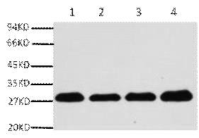
A01040-1.jpg&&Fig.1. Western blot analysis of Hela (1), rat brain (2), NIH 3T3(3), 293T(4), diluted at 1:5000.|||A01040-2.jpg&&Fig.2. Immunofluorescence analysis of human lung cancer tissue. 1, PCNA Monoclonal Antibody (1D7) (red) was diluted at 1:200 (4�C, overnight). 2, Cy3 Labeled secondary antibody was diluted at 1:300 (room temperature, 50min). 3, Picture B: DAPI (blue) 10min. Picture A: Target. Picture B: DAPI. Picture C: merge of A+B.|||A01040-3.jpg&&Fig.3. Immunofluorescence analysis of rat testis tissue. 1, PCNA Monoclonal Antibody (1D7) (red) was diluted at 1:200 (4�C, overnight). 2, Cy3 Labeled secondary antibody was diluted at 1:300 (room temperature, 50min). 3, Picture B: DAPI (blue) 10min. Picture A: Target. Picture B: DAPI. Picture C: merge of A+B.|||A01040-4.jpg&&Fig.4. Immunohistochemical analysis of paraffin-embedded human uterus tissue. 1, PCNA Monoclonal Antibody (1D7) was diluted at 1:200 (4�C, overnight). 2, Sodium citrate pH 6.0 was used for antibody retrieval(>98�C, 20min). 3, secondary antibody was diluted at 1:200 (room temperature, 30min). Negative control was used by secondary antibody only.|||A01040-5.jpg&&Fig.5. Immunohistochemical analysis of paraffin-embedded mouse liver tissue. 1, PCNA Monoclonal Antibody (1D7) was diluted at 1:200 (4�C, overnight). 2, Sodium citrate pH 6.0 was used for antibody retrieval(>98�C, 20min). 3, secondary antibody was diluted at 1:200 (room temperature, 30min). Negative control was used by secondary antibody only.|||A01040-6.jpg&&Fig.6. Immunohistochemical analysis of paraffin-embedded rat testis tissue. 1, PCNA Monoclonal Antibod y(1D7) was diluted at 1:200 (4�C, overnight). 2, Sodium citrate pH 6.0 was used for antibody retrieval(>98�C, 20min). 3, secondary antibody was diluted at 1:200 (room temperature, 30min). Negative control was used by secondary antibody only.

A01040-1.jpg&&Fig.1. Western blot analysis of Hela (1), rat brain (2), NIH 3T3(3), 293T(4), diluted at 1:5000.|||A01040-2.jpg&&Fig.2. Immunofluorescence analysis of human lung cancer tissue. 1, PCNA Monoclonal Antibody (1D7) (red) was diluted at 1:200 (4�C, overnight). 2, Cy3 Labeled secondary antibody was diluted at 1:300 (room temperature, 50min). 3, Picture B: DAPI (blue) 10min. Picture A: Target. Picture B: DAPI. Picture C: merge of A+B.|||A01040-3.jpg&&Fig.3. Immunofluorescence analysis of rat testis tissue. 1, PCNA Monoclonal Antibody (1D7) (red) was diluted at 1:200 (4�C, overnight). 2, Cy3 Labeled secondary antibody was diluted at 1:300 (room temperature, 50min). 3, Picture B: DAPI (blue) 10min. Picture A: Target. Picture B: DAPI. Picture C: merge of A+B.|||A01040-4.jpg&&Fig.4. Immunohistochemical analysis of paraffin-embedded human uterus tissue. 1, PCNA Monoclonal Antibody (1D7) was diluted at 1:200 (4�C, overnight). 2, Sodium citrate pH 6.0 was used for antibody retrieval(>98�C, 20min). 3, secondary antibody was diluted at 1:200 (room temperature, 30min). Negative control was used by secondary antibody only.|||A01040-5.jpg&&Fig.5. Immunohistochemical analysis of paraffin-embedded mouse liver tissue. 1, PCNA Monoclonal Antibody (1D7) was diluted at 1:200 (4�C, overnight). 2, Sodium citrate pH 6.0 was used for antibody retrieval(>98�C, 20min). 3, secondary antibody was diluted at 1:200 (room temperature, 30min). Negative control was used by secondary antibody only.|||A01040-6.jpg&&Fig.6. Immunohistochemical analysis of paraffin-embedded rat testis tissue. 1, PCNA Monoclonal Antibod y(1D7) was diluted at 1:200 (4�C, overnight). 2, Sodium citrate pH 6.0 was used for antibody retrieval(>98�C, 20min). 3, secondary antibody was diluted at 1:200 (room temperature, 30min). Negative control was used by secondary antibody only.