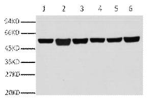
A01030-1.jpg&&Fig.1. Western blot analysis of A549(1), rat brain (2), mouse brain (3), chicken lung (4) and rabbit testis(5), sheep muscle(6), diluted at 1:10000.|||A01030-2.jpg&&Fig.2. Immunofluorescence analysis of human appendix tissue. 1, ?-Tubulin Monoclonal Antibody (3G6) (red) was diluted at 1:400 (4�C, overnight). Picture A: Target. Picture B: DAPI. Picture C: merge of A+B.|||A01030-3.jpg&&Fig.3. Immunofluorescence analysis of mouse lung tissue. 1, ?-Tubulin Monoclonal Antibody (3G6) (red) was diluted at 1:400 (4�C, overnight). Picture A: Target. Picture B: DAPI. Picture C: merge of A+B.|||A01030-4.jpg&&Fig.4. Immunohistochemical analysis of paraffin-embedded human colon tissue. 1, ?-Tubulin Monoclonal Antibody (3G6) was diluted at 1:400 (4�C, overnight). Negative control was used by secondary antibody only.|||A01030-5.jpg&&Fig.5. Immunohistochemical analysis of paraffin-embedded mouse testis tissue. 1, ?-Tubulin Monoclonal Antibody (3G6) was diluted at 1:400 (4�C, overnight). Negative control was used by secondary antibody only.|||A01030-6.jpg&&Fig.6. Immunohistochemical analysis of paraffin-embedded rat lung tissue. 1, ?-Tubulin Monoclonal Antibody (3G6) was diluted at 1:400 (4�C, overnight). Negative control was used by secondary antibody only.


