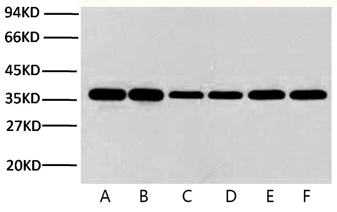
A01020-wb.jpg&&Fig.1. Western blot analysis of Hela (1), rat brain (2), rabbit muscle(3), sheep muscle(4), and mouse brain (5), diluted at 1:10000.|||A01020-2.jpg&&Fig.2. Immunohistochemical analysis of paraffin-embedded human colon tissue. 1, GAPDH Monoclonal Antibody (2B5) was diluted at 1:400 (4�C, overnight). Negative control was used by secondary antibody only.|||A01020-3.jpg&&Fig.3. Immunohistochemical analysis of paraffin-embedded mouse heart tissue. 1, GAPDH Monoclonal Antibody (2B5) was diluted at 1:400 (4�C, overnight). Negative control was used by secondary antibody only.|||A01020-4.jpg&&Fig.4. Immunohistochemical analysis of paraffin-embedded rat kidney tissue. 1, GAPDH Monoclonal Antibody (2B5) was diluted at 1:400 (4�C, overnight). Negative control was used by secondary antibody only.|||A01020-5.jpg&&Fig.5. Immunofluorescence analysis of human colon tissue. 1, GAPDH Monoclonal Antibody (2B5) (red) was diluted at 1:400 (4�C, overnight). Picture A: Target. Picture B: DAPI. Picture C: merge of A+B.|||A01020-6.jpg&&Fig.6. Immunofluorescence analysis of mouse liver tissue. 1, GAPDH Monoclonal Antibody (2B5) (red) was diluted at 1:400 (4�C, overnight). Picture A: Target. Picture B: DAPI. Picture C: merge of A+B.|||A01020-7.jpg&&Fig.7 Western blot analysis of Pig muscle tissue, GAPDH Monoclonal Antibody (2B5) was diluted at 1:10000 (25�C, 3h).|||A01020-8.jpg&&Fig.8 Western blot analysis of Hela, GAPDH Monoclonal Antibody (2B5) was diluted at 1:10000 (25�C, 3h).


