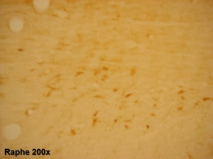
IHC image of neurons in the rat basal forebrain staining for VAT. The tissue was fixed with 4% formaldehyde in phosphate buffer, before being removed and prepared for vibratome sectioning. Floating sections were incubated at RT in 10% rabbit serum in PBS, before standard IHC procedure. Primary antibody was incubated at 1:5000 for 48 hours, rabbit anti-goat secondary was subsequently added for 1 hour after washing with PBS. Light microscopy staining was achieved with standard biotin-streptavidin/HRP procedure and DAB chromogen.


