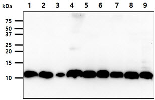
The cell lysates (40ug) were resolved by SDS-PAGE, transferred to PVDF membrane and probed with anti-human S100A11 antibody (1:1000). Proteins were visualized using a goat anti-mouse secondary antibody conjugated to HRP and an ECL detection system.Lane 1. : HeLa cell lysate Lane 2. : MCF7 cell lysate Lane 3. : PC3 cell lysate Lane 4. : HaCaT cell lysate Lane 5. : THP1 cell lysate Lane 6. : U87MG cell lysate Lane 7. : A427 cell lysate Lane 8. : SK-OV-3 cell lysate Lane 9. : A549 cell lysate
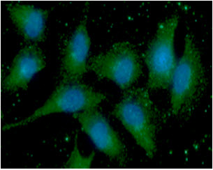
ICC/IF analysis of S100A11 in A549 cells line, stained with DAPI (Blue) for nucleus staining and monoclonal anti-human S100A11 antibody (1:100) with goat anti-mouse IgG-Alexa fluor 488 conjugate (Green).


