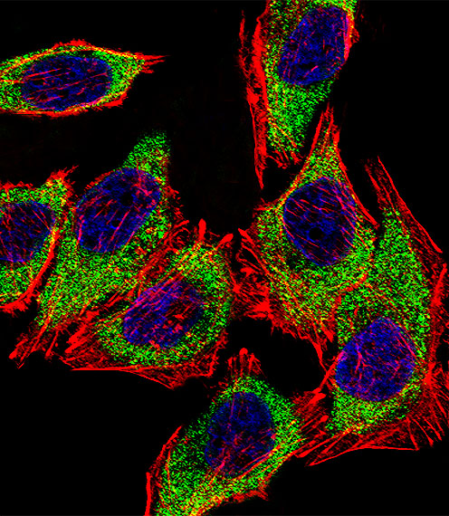
Fluorescent confocal image of A549 cell stained with AGR2 Antibody (N-term)(Cat#AP6279a).A549 cells were fixed with 4% PFA (20 min), permeabilized with Triton X-100 (0.1%, 10 min), then incubated with AGR2 primary antibody (1:25, 1 h at 37?). For secondary antibody, Alexa Fluor� 488 conjugated donkey anti-rabbit antibody (green) was used (1:400, 50 min at 37?).Cytoplasmic actin was counterstained with Alexa Fluor� 555 (red) conjugated Phalloidin (7units/ml, 1 h at 37?). Nuclei were counterstained with DAPI (blue) (10 �g/ml, 10 min). AGR2 immunoreactivity is localized to Cytoplasm significantly.


