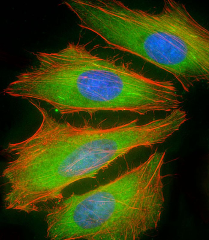
Immunofluorescent analysis of 4% paraformaldehyde-fixed, 0.1% Triton X-100 permeabilized HeLa (human cervical epithelial adenocarcinoma cell line) cells labeling BCL2L10 with AP22305c at 1/25 dilution, followed by Dylight� 488-conjugated goat anti-rabbit IgG (NK179883) secondary antibody at 1/200 dilution (green). Immunofluorescence image showing cytoplasm and nucleus staining on HeLa cell line. Cytoplasmic actin is detected with Dylight� 554 Phalloidin (PD18466410) at 1/100 dilution (red). The nuclear counter stain is DAPI (blue).


