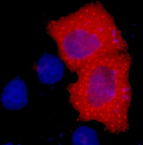
A02040-if.jpg&&Fig.1. Immunofluorescence staining (1:2000) of HA fusion protein in 293 cells with red and counterstained with DAPI.|||A02040-wb.jpg&&Fig.2.IP (1:400)-WB (1:10000) analysis of HA fusion protein expression in 293 cells. Untransfected 293 cell lysate (lane A), transfected 293 cell lysate with HA-tag protein (lane B); IP untransfected 293 cell lysate with Anti HA tag mAb (lane C); IP transfected 293 cell lysate with normal Mouse IgG (lane D), or with Anti HA tag mAb (lane E), and IP transfected 293 without both normal Mouse IgG and HA tag mAb (lane F).


